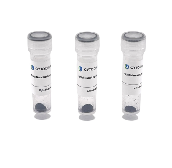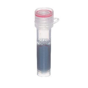
Lateral Flow Assay Product - 015nm 10nm
Lateral Flow Assay Product - 015nm 10nm for diagnostic research and development.
Compare products with technical highlights, document cues, and product details.

Lateral Flow Assay Product - 10nm for diagnostic research and development.

Lateral Flow Assay Product - 50nm for diagnostic research and development.

Membrane & Assay Component (MA-MFI-001) for diagnostic research and development.

Membrane & Assay Component (MA-MFI-002) for diagnostic research and development.

MWCO 10 kDa Ultrafiltration Spin Column for diagnostic research and development.

MWCO 100 kDa Ultrafiltration Spin Column for diagnostic research and development.

MWCO 30 kDa Ultrafiltration Spin Column for diagnostic research and development.

MWCO 5 kDa Ultrafiltration Spin Column for diagnostic research and development.

Product Description Sample cleanup, desalting, concentrating centrifugal filters. Simple one-spin function for rapid concentration of samples, and removal of small contaminants (salt, dyes, radioactive labels). Polyethersulfone (PES) membrane for maximum recovery of protein and DNA. Ultrafiltration spin units offer a simple, one-step procedure for desalting and concentration of proteins and other macromolecules with greater than 90% recovery. Utilizing a molecular weight cut-off polyethersulfone (PES) filter membrane, the spin device is able to retain macromolecules having a greater MW, and filtrate salts and other molecules of smaller MW. Because of low protein/DNA binding properties of the PES filter, loss of the retentate on the filter is minimized. The retentate can be recovered at excellent rate. Four molecular weight cut-off (MWCO) sizes are provided to cover a broad range of molecular weights: 5 kDa, 10 kDa, 30 kDa, and 100 kDa. It is recommended to choose a MWCO 1/2 to 1/3 smaller than the protein to be concentrated. For instance, to concentrate a protein of 80 kDa, we recommend to start with the 30 kDa MWCO spin unit.

MWCO Ultrafiltration Spin Columns, 10 kDa- 5 columns for diagnostic research and development.

Product Description Sample cleanup, desalting, concentrating centrifugal filters. Simple one-spin function for rapid concentration of samples, and removal of small contaminants (salt, dyes, radioactive labels). Polyethersulfone (PES) membrane for maximum recovery of protein and DNA. Ultrafiltration spin units offer a simple, one-step procedure for desalting and concentration of proteins and other macromolecules with greater than 90% recovery. Utilizing a molecular weight cut-off polyethersulfone (PES) filter membrane, the spin device is able to retain macromolecules having a greater MW, and filtrate salts and other molecules of smaller MW. Because of low protein/DNA binding properties of the PES filter, loss of the retentate on the filter is minimized. The retentate can be recovered at excellent rate. Four molecular weight cut-off (MWCO) sizes are provided to cover a broad range of molecular weights: 5 kDa, 10 kDa, 30 kDa, and 100 kDa. It is recommended to choose a MWCO 1/2 to 1/3 smaller than the protein to be concentrated. For instance, to concentrate a protein of 80 kDa, we recommend to start with the 30 kDa MWCO spin unit.

MWCO Ultrafiltration Spin Columns, 100 kDa- 5 columns for diagnostic research and development.

Product Description Sample cleanup, desalting, concentrating centrifugal filters. Simple one-spin function for rapid concentration of samples, and removal of small contaminants (salt, dyes, radioactive labels). Polyethersulfone (PES) membrane for maximum recovery of protein and DNA. Ultrafiltration spin units offer a simple, one-step procedure for desalting and concentration of proteins and other macromolecules with greater than 90% recovery. Utilizing a molecular weight cut-off polyethersulfone (PES) filter membrane, the spin device is able to retain macromolecules having a greater MW, and filtrate salts and other molecules of smaller MW. Because of low protein/DNA binding properties of the PES filter, loss of the retentate on the filter is minimized. The retentate can be recovered at excellent rate. Four molecular weight cut-off (MWCO) sizes are provided to cover a broad range of molecular weights: 5 kDa, 10 kDa, 30 kDa, and 100 kDa. It is recommended to choose a MWCO 1/2 to 1/3 smaller than the protein to be concentrated. For instance, to concentrate a protein of 80 kDa, we recommend to start with the 30 kDa MWCO spin unit.

MWCO Ultrafiltration Spin Columns, 30 kDa- 5 columns for diagnostic research and development.

Product Description Sample cleanup, desalting, concentrating centrifugal filters. Simple one-spin function for rapid concentration of samples, and removal of small contaminants (salt, dyes, radioactive labels). Polyethersulfone (PES) membrane for maximum recovery of protein and DNA. Ultrafiltration spin units offer a simple, one-step procedure for desalting and concentration of proteins and other macromolecules with greater than 90% recovery. Utilizing a molecular weight cut-off polyethersulfone (PES) filter membrane, the spin device is able to retain macromolecules having a greater MW, and filtrate salts and other molecules of smaller MW. Because of low protein/DNA binding properties of the PES filter, loss of the retentate on the filter is minimized. The retentate can be recovered at excellent rate. Four molecular weight cut-off (MWCO) sizes are provided to cover a broad range of molecular weights: 5 kDa, 10 kDa, 30 kDa, and 100 kDa. It is recommended to choose a MWCO 1/2 to 1/3 smaller than the protein to be concentrated. For instance, to concentrate a protein of 80 kDa, we recommend to start with the 30 kDa MWCO spin unit.

MWCO Ultrafiltration Spin Columns, 5 kDa- 5 columns for diagnostic research and development.

Designed for researchers, the Miriad® product line delivers versatile, Research Use Only solutions for multi-marker screening across blood banks, tissue and eye banks, organ donor programs, and scientific studies. Available worldwide without regulatory barriers, Miriad® offers ready-made and tailored combinations for detecting multiple disease markers. Built on our RVF Technology™, these rapid antibody tests provide immediate, easy-to-interpret results that enhance research and support informed decision-making in donor evaluation and advanced study environments. Immediate results - No waiting time, results do not vanish. Reliable accuracy - Achieves high testing performance across multiple specimen matrices. Multiplex testing - Multiple diseases tested on a single cartridge using one specimen. No equipment or refrigeration required - Stores at room temperature (2–30 °C / 35–86 °F) with a 18-month shelf life. Easy to use and interpret -Simple test procedure with easy interpretation, customer support is offered for development. For Research Use Only - Works with serum, plasma, and venipuncture blood.

Designed for researchers, the Miriad® product line delivers versatile, Research Use Only solutions for multi-marker screening across blood banks, tissue and eye banks, organ donor programs, and scientific studies. Available worldwide without regulatory barriers, Miriad® offers ready-made and tailored combinations for detecting multiple disease markers. Built on our RVF Technology™, these rapid antibody tests provide immediate, easy-to-interpret results that enhance research and support informed decision-making in donor evaluation and advanced study environments. Immediate results - No waiting time, results do not vanish. Reliable accuracy - Achieves high testing performance across multiple specimen matrices. Multiplex testing - Multiple diseases tested on a single cartridge using one specimen. No equipment or refrigeration required - Stores at room temperature (2–30 °C / 35–86 °F) with a 18-month shelf life. Easy to use and interpret -Simple test procedure with easy interpretation, customer support is offered for development. For Research Use Only - Works with serum, plasma, and venipuncture blood.

Kit Components 10 Miriad™ test cartridges 12 ml of universal buffer 20nm Protein A Gold Conjugate (2 ml, OD10) Detailed product guide To detect antibodies using this toolkit the selected antigen(s) can be spotted onto the nitrocellulose membrane encapsulated within the specially designed plastic test cartridge. When the sample is applied any antibodies against the immobilized antigens in the sample will form an antigen/antibody immuno-complex, which is detected through the use of the supplied Protein A-Gold Conjugate. Up to five antigens can be spotted onto one cartridge making this kit ideal for screening of antibody specificity.

Kit Components 10 Miriad™ test cartridges 12 ml of universal buffer 80nm Protein A Gold NanoUrchin Conjugate (2 ml, OD10) Detailed product guide To detect antibodies using this toolkit the selected antigen(s) can be spotted onto the nitrocellulose membrane encapsulated within the specially designed plastic test cartridge. When the sample is applied any antibodies against the immobilized antigens in the sample will form an antigen/antibody immuno-complex, which is detected through the use of the supplied Protein A-Gold NanoUrchin Conjugate which gives a blue spot upon positive detection. Up to five antigens can be spotted onto one cartridge making this kit ideal for screening of antibody specificity.

Kit Components 10 Miriad™ test cartridges 12 ml of universal buffer Lyophilized 20nm gold nanoparticles (for conjugation to your primary antibody) Protein Resuspension Buffer Reaction Buffer Quencher Detailed product guide with conjugation and assay procedures Required reagents not included in kit Primary antibody/antibodies - For antigen capture on the membrane and conjugation to gold nanoparticles (detection probe). Assay Principle To detect an antigen using this toolkit a primary antibody is spotted onto the nitrocellulose membrane encapsulated within the specially designed plastic test cartridge. When the sample is applied to the cartridge any antigen present will bind to the capture antibody immobilized on the membrane. This binding event is subsequently detected by the formation of a sandwich through the binding of a gold conjugated primary antibody to the bound antigen.A positive sample will be indicated by the formation of a red spot on the membrane, figure 1. https://cdn.shopify.com/s/files/1/0256/1107/5658/files/Positive-vs-Negative-Vertical-Flow-Sample.jpg?v=1573662133.

Kit Components 10 Miriad™ test cartridges 12 ml of universal buffer Lyophilized 80nm Gold NanoUrchins (for conjugation to your primary antibody) Protein Resuspension Buffer Reaction Buffer Quencher Detailed product guide with conjugation and assay procedures Required reagents not included in kit Primary antibody/antibodies - For antigen capture on the membrane and conjugation to gold nanoparticles (detection probe). Assay Principle To detect an antigen using this toolkit a primary antibody is spotted onto the nitrocellulose membrane encapsulated within the specially designed plastic test cartridge. When the sample is applied to the cartridge any antigen present will bind to the capture antibody immobilized on the membrane. This binding event is subsequently detected by the formation of a sandwich through the binding of a Gold NanoUrchin conjugated primary antibody to the bound antigen. A positive sample will be indicated by the formation of a blue spot on the membrane, figure 1. https://cdn.shopify.com/s/files/1/0256/1107/5658/files/Positive-vs-Negative-Vertical-Flow-Sample.jpg?v=1573662133.

Kit Components 10 Miriad™ test cartridges / 50 Miriad™ test cartridges 12 ml of universal buffer / 60 ml of universal buffer Detailed product guide To detect antibodies using this toolkit the selected antigen(s) can be spotted onto the nitrocellulose membrane encapsulated within the specially designed plastic test cartridge. When the sample is applied any antibodies against the immobilized antigens in the sample will form an antigen/antibody immuno-complex, which is detected through the use of a Protein A-, or a Protein G-gold conjugate (not supplied with this kit). The kit is also suitable to detect specific antigen molecules. In this case a pair of antibodies, one for capturing the antigen (spotted onto the membrane as above) and the other for conjugation to gold nanoparticles for detection will be required. When the sample is applied the spotted antibody will capture any target antigen present, which can subsequently be visualized by applying the detection antibody-gold conjugate.