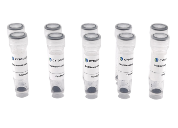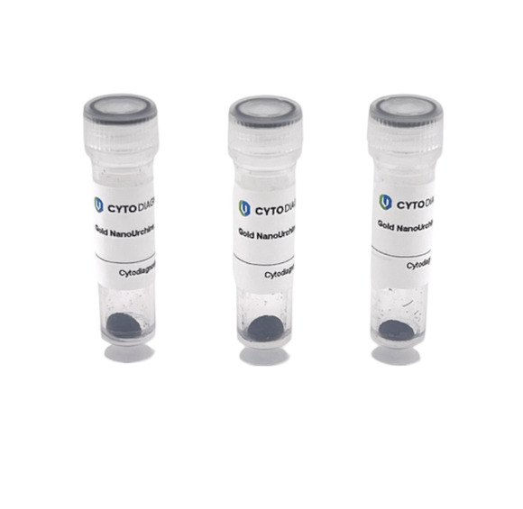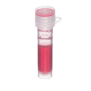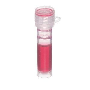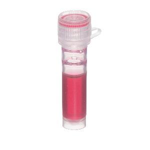
Gold NanoUrchin Conjugate - 90nm 20nm 05nm
Gold NanoUrchin Conjugate - 90nm 20nm 05nm for diagnostic research and development.
Compare products with technical highlights, document cues, and product details.

Gold Nanoparticle Conjugate - 100nm 05nm for diagnostic research and development.

Gold Nanoparticle Conjugate - 10nm 05nm for diagnostic research and development.

Gold Nanoparticle Conjugate - 15nm 05nm for diagnostic research and development.

Gold Nanoparticle Conjugate - 20nm 05nm for diagnostic research and development.

Gold Nanoparticle Conjugate - 30nm 05nm for diagnostic research and development.

Gold Nanoparticle Conjugate - 40nm 05nm for diagnostic research and development.

Gold Nanoparticle Conjugate - 50nm 05nm for diagnostic research and development.

Gold Nanoparticle Conjugate - 5nm 05nm for diagnostic research and development.

Gold Nanoparticle Conjugate - 60nm 05nm for diagnostic research and development.

Gold Nanoparticle Conjugate - 80nm 05nm for diagnostic research and development.

Gold Nanoparticle Conjugate - 90nm 05nm for diagnostic research and development.

Supplied with Kit 20nm and 30nm standard gold nanoparticles (100ml each, conc. OD1) 12 conjugation optimization buffers (1.5ml each) 10% NaCl (100ml) Protein resuspension buffer Detailed conjugation optimization protocol Kit contains enough material to optimize 10 protein conjugates, or one optimization reaction and scale-up production of 20ml of conjugate at a final optical density of OD3. Note: Only use with Cytodiagnostics standard or reactant free gold nanoparticles. Gold nanoparticle conjugates have been used for a wide range of biological applications including the use as probes in light and electron microscopy (TEM) and for membrane blotting (immunogold dot-blot). Stable gold nanoparticle conjugates can readily be prepared by passive adsorption of a protein to the gold nanoparticle surface, which is mediated by electrostatic and hydrophobic interactions between the protein and the surface layer of the colloidal gold. Our gold nanoparticle protein conjugation optimization kits are supplied as complete and easy to use systems for increased success rates in the development of good quality gold nanoparticle conjugates.

Supplied in Kit: 500ml Ready to use Gold Membrane Stain 100ml Wash Buffer (100X) Cytodiagnostics Gold Protein Detection Kit for Membranes is a stabilized gold solution specially developed to stain proteins blotted onto nitrocellulose or polyvinylidene difluoride (PVDF) membranes. The negatively charged gold particles bind to protein(s) on the membrane through ionic and hydrophobic interaction with much higher affinity than to the membrane itself. Accumulation of gold particles onto the bound proteins produces dark permanent red bands, with better sensitivity (down to 1 ng, protein dependent). The kit is provided ready to use; no reconstitution or dilution required. When used in conjunction with Cytodiagnostics Silver Enhancement Kit for Membranes (Cat. # SR-01-02), a further improvement in sensitivity (10 to 100 times) can be acheived. Storage This product should be stored at 4°C and is stable for at least 6 months. Do not freeze.

Lateral Flow Assay Product (LF-006-25) for diagnostic research and development.

upplied with Kit 50nm standard GoldNanourchins (100ml, conc. OD1) 12 conjugation optimization buffers (1.5ml each) 10% NaCl (100ml) Protein resuspension buffer Detailed conjugation optimization protocol Kit contains enough material to optimize 10 protein conjugates, or one optimization reaction and scale-up production of 20ml of conjugate at a final optical density of OD3. Gold NanoUrchin conjugates have been used for a wide range of biological applications including the use as probes in light and electron microscopy (TEM) and for membrane blotting (immunogold dot-blot); especially where large surface plasmon resonance shifts are required. Stable Gold NanoUrchin conjugates can readily be prepared by passive adsorption of a protein to the Gold NanoUrchin surface, which is mediated by electrostatic and hydrophobic interactions between the protein and the surface layer of the colloidal gold. Our Gold NanoUrchin protein conjugation optimization kits are supplied as complete and easy to use systems for increased success rates in the development of good quality Gold NanoUrchin conjugates.

Supplied with Kit 80nm standard GoldNanourchins (100ml, conc. OD1) 12 conjugation optimization buffers (1.5ml each) 10% NaCl (100ml) Protein resuspension buffer Detailed conjugation optimization protocol Kit contains enough material to optimize 10 protein conjugates, or one optimization reaction and scale-up production of 20ml of conjugate at a final optical density of OD3. Gold NanoUrchin conjugates have been used for a wide range of biological applications including the use as probes in light and electron microscopy (TEM) and for membrane blotting (immunogold dot-blot); especially where large surface plasmon resonance shifts are required. Stable Gold NanoUrchin conjugates can readily be prepared by passive adsorption of a protein to the Gold NanoUrchin surface, which is mediated by electrostatic and hydrophobic interactions between the protein and the surface layer of the colloidal gold. Our Gold NanoUrchin protein conjugation optimization kits are supplied as complete and easy to use systems for increased success rates in the development of good quality Gold NanoUrchin conjugates.

Supplied with Kit 20nm standard gold nanoparticles (100ml, conc. OD1) 12 conjugation optimization buffers (1.5ml each) 10% NaCl (100ml) Protein resuspension buffer Detailed conjugation optimization protocol Kit contains enough material to optimize 10 protein conjugates, or one optimization reaction and scale-up production of 20ml of conjugate at a final optical density of OD3. Note: Only use with Cytodiagnostics standard or reactant free gold nanoparticles. Gold nanoparticle conjugates have been used for a wide range of biological applications including the use as probes in light and electron microscopy (TEM) and for membrane blotting (immunogold dot-blot). Stable gold nanoparticle conjugates can readily be prepared by passive adsorption of a protein to the gold nanoparticle surface, which is mediated by electrostatic and hydrophobic interactions between the protein and the surface layer of the colloidal gold. Our gold nanoparticle protein conjugation optimization kits are supplied as complete and easy to use systems for increased success rates in the development of good quality gold nanoparticle conjugates.

Supplied with Kit 40nm standard gold nanoparticles (100ml, conc. OD1) 12 conjugation optimization buffers (1.5ml each) 10% NaCl (100ml) Protein resuspension buffer Detailed conjugation optimization protocol Kit contains enough material to optimize 10 protein conjugates, or one optimization reaction and scale-up production of 20ml of conjugate at a final optical density of OD3. Note: Only use with Cytodiagnostics standard or reactant free gold nanoparticles. Gold nanoparticle conjugates have been used for a wide range of biological applications including the use as probes in light and electron microscopy (TEM) and for membrane blotting (immunogold dot-blot). Stable gold nanoparticle conjugates can readily be prepared by passive adsorption of a protein to the gold nanoparticle surface, which is mediated by electrostatic and hydrophobic interactions between the protein and the surface layer of the colloidal gold. Our gold nanoparticle protein conjugation optimization kits are supplied as complete and easy to use systems for increased success rates in the development of good quality gold nanoparticle conjugates.

Supplied with Kit 60nm standard gold nanoparticles (100ml, conc. OD1) 12 conjugation optimization buffers (1.5ml each) 10% NaCl (100ml) Protein resuspension buffer Detailed conjugation optimization protocol Kit contains enough material to optimize 10 protein conjugates, or one optimization reaction and scale-up production of 20ml of conjugate at a final optical density of OD3. Note: Only use with Cytodiagnostics standard or reactant free gold nanoparticles. Gold nanoparticle conjugates have been used for a wide range of biological applications including the use as probes in light and electron microscopy (TEM) and for membrane blotting (immunogold dot-blot). Stable gold nanoparticle conjugates can readily be prepared by passive adsorption of a protein to the gold nanoparticle surface, which is mediated by electrostatic and hydrophobic interactions between the protein and the surface layer of the colloidal gold. Our gold nanoparticle protein conjugation optimization kits are supplied as complete and easy to use systems for increased success rates in the development of good quality gold nanoparticle conjugates.

40nm and 60nm standard gold nanoparticles (100ml each, conc. OD1) 12 conjugation optimization buffers (1.5ml each) 10% NaCl (100ml) Protein resuspension buffer Detailed conjugation optimization protocol Kit contains enough material to optimize 10 protein conjugates, or one optimization reaction and scale-up production of 20ml of conjugate at a final optical density of OD3.

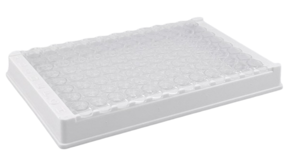
Product overview: The interleukin (IL)-17 family comprises six members: IL-17A, IL-17B, IL-17C, IL-17D, IL-17E, and IL-17F, while IL-17A is the most studied subtype. Human IL-17A is a 15-20 kDa glycosylated polypeptide, a proinflammatory cytokine primarily produced by T helper 17 (Th17) cells, alongside γδ T cells, natural killer T cells, and innate lymphoid cells. IL-17A forms both disulfide-linked homodimers (32-38 kDa) and covalent heterodimers with IL-17F (40-45 kDa). While most secreted IL-17A exists as the IL-17A:F heterodimer, the IL-17A:A homodimer is more bioactive. IL-17 plays a critical role in immune responses against extracellular pathogens and contributes to the pathogenesis of various autoimmune and inflammatory diseases. IL-17A has attracted significant attention in scientific research due to its regulatory role in immune and inflammatory processes. IL-17 induces neutrophil recruitment and stimulates the production of pro-inflammatory cytokines like IL-6, IL-8, TNF-α, and IL-1β. Elevated IL-17A levels are associated with several chronic inflammatory diseases in humans, including rheumatoid arthritis, asthma, acute coronary syndrome, multiple sclerosis, systemic lupus erythematosus, and allograft rejection. Our ELISA kit has several benefits: Ready to use. Simply add standards and samples directly to wells. No need for lengthy blocking or rehydration steps. High sensitivity, specificity and reproducibility. Fully validated in the sample types listed below. 96-well plate is breakable into 12 x 8 well strips.

Product overview: Interleukin-6 (IL-6), a 26 kDa glycoprotein composing of 184 amino acids, plays a pivotal role in inflammation, immune responses, hematopoiesis, and oncogenesis. While its typical circulating level in healthy adults is less than 5 pg/mL, they escalate rapidly during inflammation. IL-6 is expressed in diverse cell types, including adipocytes, fibroblasts, osteoblasts, neurons, and immune cells. Elevated IL-6 levels are associated with conditions like cardiovascular disease, insulin resistance, type 2 diabetes, asthma, rheumatoid arthritis, and advanced-stage cancer. As a signaling molecule, IL-6 regulates cell growth and differentiation, particularly in immune responses and acute phase reactions. Secreted by various cell types, including T cells and macrophages, IL-6 exerts its actions through a heterodimeric receptor composed of IL-6R and glycoprotein 130 (gp130). This receptor complex transduces signals, contributing to immune responses, hematopoiesis, bone metabolism, and cancer progression. IL-6 is not constitutively produced but is induced by factors like cytokines, lipopolysaccharide, or viral infections. Its broad tissue effects vary from inducing growth and inhibition to differentiation, depending on the target cells. Abnormal IL-6 production is implicated in various diseases, and measuring its levels in serum provides diagnostic insights. The multifaceted nature of IL-6 and its involvement in diverse physiological processes make it a central figure in immunology, with significant implications for both research and clinical applications. Our ELISA kit has several benefits: Ready to use. Simply add standards and samples directly to wells. No need for lengthy blocking or rehydration steps. High sensitivity, specificity and reproducibility. Fully validated in the sample types listed below. 96-well plate is breakable into 12 x 8 well strips.